Chemical Nano-Characterization by Correlative Analyzes in Electron Microscopy and Secondary Ions Mass Spectroscopy: Example on Ni-Based Superalloys
Secondary-ion mass spectrometry (SIMS) is probably the most widely used chemical analysis technique in semiconductor science and in metallurgy. Due to its ultimate sensitivity to all elements and in particular, to light elements, this technology provides semi-quantitative information on the depth distribution of elements, for instance, doping elements (i.e., P, B, H) or contaminants (i.e., C and O). SIMS also provides information on the chemical gradients, and more generally on the chemical information in 2D or 3D, for instance, segregation in thin films and at interfaces. The development of an innovative analytical SIMS implemented in a NanoSpace platform with a focused Gallium source ion beam (FIB) and a scanning electron microscope (SEM) enables to give elemental chemical mapping at very high resolution (<30nm) and high mass resolution (4,500 on 28Si). This instrument allows correlative SEM/SIMS analysis in the same instrument and SIMS analysis can be performed on a specific region of interest revealed by secondary electron or backscattered electron non-destructive imaging.
Why using SIMS for chemical analysis
The secondary-ion mass spectrometry (SIMS) method is commonly used to detect trace elements and light elements due to its ultimate sensitivity. In addition, the ability to dissociate isotopes is useful in many applications as the isotope ratio can provide different information about materials. For example, tracer O will give insight into oxidation or 2H tracer on mechanisms of hydrogenation in metallic superalloys. Here, the technique will allow to characterize at the nanoscale the Ni-based superalloys used in the manufacturing of engine parts and accessories for aircraft and aerospace equipment. They are designed for their high mechanical and thermal resistance. The time-of-flight (TOF)-SIMS is a well-known technique where a primary ion beam sputters the sample locally and where a part of the sputtered material is ionized, forming the secondary ions.This ionization rate is then strongly dependent of the considered element and of the chemical composition of the sample. They both influence the number of secondary ions collected at each point of the sample and then the chemical distribution maps. Once collected, those ions are transmitted to a mass spectrometer which measures their time of flight. Our solution, the Orthogonal TOF- SIMS used here, allows a continuous sputtering of the material and a pulsation of the secondary ion beam. This approach allows to keep all benefits of high spatial resolution from the FIB without losing mass resolution.
Experimental conditions
The SIMS measurements were performed using the standalone Orsay Physics instrument, named NanoSpace. In order to obtain the best high spatial resolution and high mass resolution, the platform was equipped with a FIB column with an LMIS Gallium source, called Cobra, and the ExOTOF, which combines an O-TOF from Tofwerk and our own new extraction system. To locate precisely the area of interest, the NanoSpace was also equipped with an SEM column, an Eclipse Plus. A Ni-based superalloy sample was characterized due to its interest in its great diversity of precipitates in terms of size and chemical compositions. Here the EDS maps were not precise enough and the SIMS is bringing a high-resolution analysis. In this example, the elements of interest have better positive than negative ionization, therefore we have focused our study on positive secondary ions. The SIMS analyses were obtained using a 30keV primary ion energy and a probe current of 1pA, both to reduce the chromatic aberration and maximize the lateral resolution. The 2D surface analyses were obtained by scanning 15μm2, a single pass of 600x 1,000 pixels with 50 μs per pixel. The total analysis time was 30s per maps.
High-resolution chemical characterization
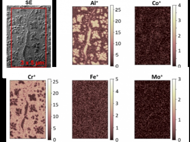
After selecting the peaks of interest in the total mass spectrum, the 2D distribution of each of these masses in the analyzed area is obtained. These chemical maps of positive secondary ions reveal a preference for the segregation of aluminum, nickel, and titanium in the precipitates, and conversely, chromium segregates towards the matrix. For the other elements, their low secondary ionization rates and/or their low concentrations give little or no imaging information. Indeed, metals like Al, Cr, and Ti have a higher ionization rate than Co, Ni, Nb, and Mo (in positive polarity). However, this analysis is qualitative, because the intensity of each element does not depend only on their concentration but also on their secondary ionization rate. It remains possible to have semiquantitative analysis to quantify their concentration from SIMS data with a calibration sample. Indeed, there is no sufficiently reliable theoretical model to predict the ionization yield of a species in a given chemical matrix.
High-resolution spatial characterization
Using an image with enough contrast, like the positive aluminum ions, very small precipitates can be observed and used to cha- racterize the spatial detection limit. To determine the spatial resolution, the measurements in 16/84 were performed on different edges of precipitates. The intensity profiles show that the SIMS detects precipitates smaller than 30nm, with an average of 25nm.
Reference:
[1] Almoric, J., Durand, M., Seret, A., Nicolaÿ, A., Houel, A., Berbezier, I. and Bozzolo, N. (2022), Implementation of Nanoscale Secondary-Ion Mass Spectrometry Analyses: Application to Ni-Based Superalloys. Phys. Status Solidi A 2100414.
[2] https://www.cemef.minesparis.psl.eu/en/presentation/metallurgie-structure-rheology-msr/
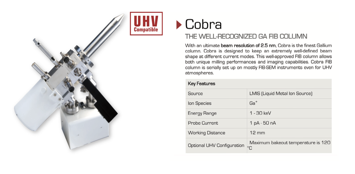
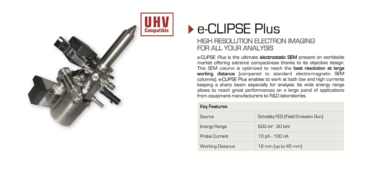
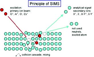
Figure 1. Schematic SIMS principle
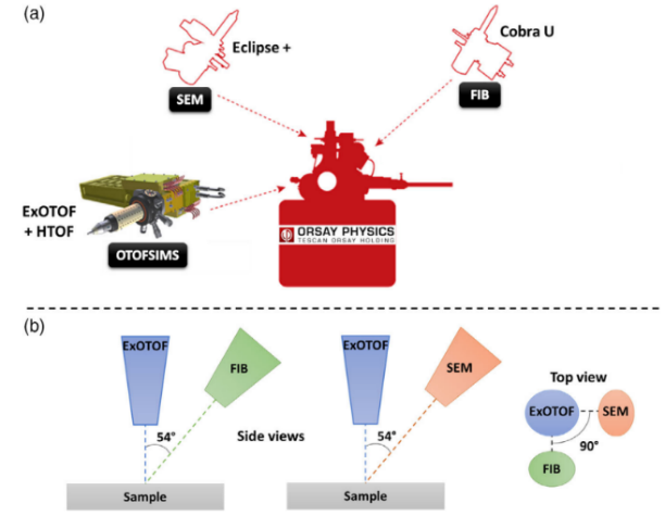
Figure 2. (a)NanoSpace configuration used for this work and made by ORSAY PHYSICS and (b) schematic views of the different positions and angles of the optic columns.
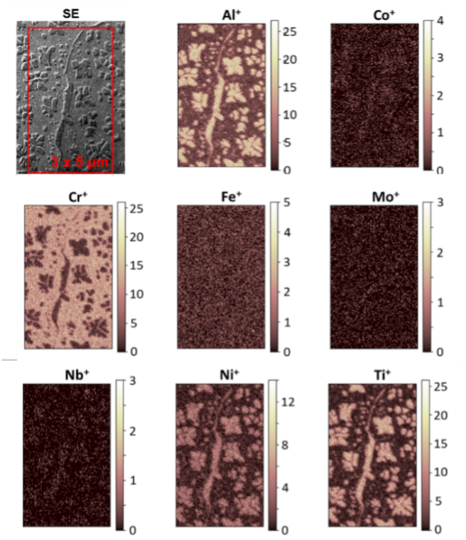
Figure 3. Chemical maps of positive secondary ions (Al+, Co+, Cr+, Fe+, Mo+, Nb+, Ni+,Ti+, and the corresponding SE image (red rectangle).